CD146 antibody | OJ79c


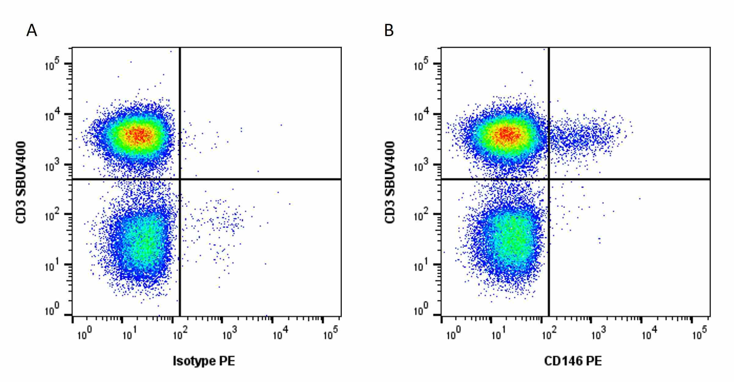
Mouse anti Human CD146 antibody, clone OJ79c (MCA2141) used for the evaluation of CD146 expression on brain derived cell lines by flow cytometry.
Image caption:
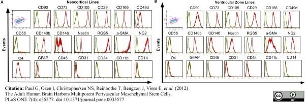
Brain-derived cell lines express markers for mesenchymal stem cells and pericytes but not for glial or neuronal precursors. (A) Representative histogram of flow cytometry analysis of cortical line and (B) ventricular zone line. Progenitors from all donors and both regions highly express MSC (CD90, CD73, CD105, CD29, CD166 and CD49d) and pericyte markers (CD140b/PDGFR-β, RGS5, CD146, Nestin, α-SMA and NG2). They do not express hematopoietic (CD45), endothelial (CD31, CD34), microglial (CD14, CD11), glial or neuronal precursor cell markers (GFAP, O4) or myofibroblast markers (CD56), (green = isotype, red = respective marker).
From: Paul G, Özen I, Christophersen NS, Reinbothe T, Bengzon J, et al. (2012)
The Adult Human Brain Harbors Multipotent Perivascular Mesenchymal Stem Cells.
PLoS ONE 7(4): e35577.
This is from an open access article distributed under the terms of the Creative Commons Attribution License.
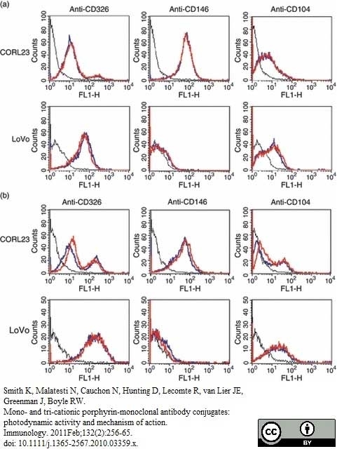
Mouse anti Human CD146 antibody, clone OJ79c (MCA2141) used for the evaluation of CD146 expression on cell lines and compared to binding of porphyrin conjugated antibodies by flow cytometry.
Image caption
Conjugated and unconjugated anti-CD326, anti-CD146 and anti-CD104 were analysed by flow cytometry for binding to CORL23 and LoVo cells [black line, negative control; blue line, unconjugated antibody; red line, immunoconjugates with porphyrin 1 (a) or porphyrin 2 (b)].
From: Smith K, Malatesti N, Cauchon N, Hunting D, Lecomte R, van Lier JE, Greenman J, Boyle RW.
Mono- and tri-cationic porphyrin-monoclonal antibody conjugates: photodynamic activity and mechanism of action.
Immunology. 2011 Feb;132(2):256-65.
This is from an open access article distributed under the terms of the Creative Commons Attribution License.
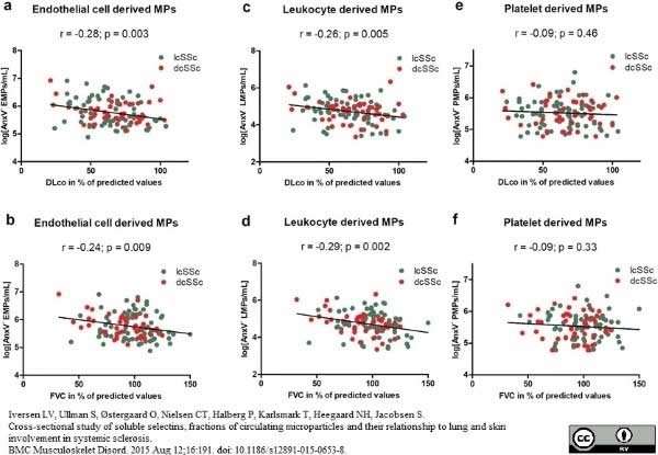
Mouse anti HumanCD146 antibody, clone OJ79c (MCA2141) used for the identification of endothelial cell derived non-annexin V binding, circulating microparticles by flow cytometry.
Image caption:
Plasma levels of annexin V non-binding, cell-derived microparticles (AnxV− MPs) correlated to lung function parameters. Endothelial cell derived MPs and leukocyte derived MPs (log counts/mL) correlated with both carbon monoxide diffusing capacity in percent of predicted values (DLco, %) (r = -0.28; p = 0.003, and r = -0.26; p = 0.005) respectively (a and b) and forced vital capacity in percent of predicted values (FVC, %) (r = -0.24; p = 0.009, and r = -0.29; p = 0.002) respectively (c and d). No correlations between AnxV− platelet derived MPs and lung function parameters were found (e and f). Correlation analysis was performed by use of Pearson's r test using log transformed microparticle data.
From: Iversen LV, Ullman S, Østergaard O, Nielsen CT, Halberg P, Karlsmark T, Heegaard NH, Jacobsen S.
Cross-sectional study of soluble selectins, fractions of circulating microparticles and their relationship to lung and skin involvement in systemic sclerosis.
BMC Musculoskelet Disord. 2015 Aug 12;16:191.
This is from an open access article distributed under the terms of the Creative Commons Attribution License.
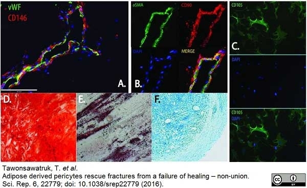
AlexaFluor 647 conjugated Mouse anti Human CD146 antibody, clone OF79c (MCA2141A647) used for the detection of CD146 expressing cells by immunofluorescence.
Image caption:
(A) Pericytes expressing CD146 reside on the abluminal surface of endothelial cells expressing vWF. (B) αSMA expressing pericytes co-express MSC markers including CD90. (C) In-vitro pericytes express MSC markers including CD105. Cultured pericytes are multipotent as seen by their ability to differentiate into bone (D) (Alizarin red staining ×10), fat (E) (Oil Red O staining × 10), cartilage (F) (Alcian blue staining × 5).
From: Tawonsawatruk, T. et al. Adipose derived pericytes rescue fractures from a failure of healing – non-union. Sci. Rep. 6, 22779.
This is from an open access article distributed under the terms of the Creative Commons Attribution License.
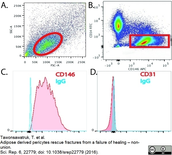
AlexaFluor 647 conjugated Mouse anti Human CD146 antibody, clone OF79c (MCA2141A647) used for the detection of CD146 expressing cells by flow cytometry.
Image caption:
(A) Pericytes can be purified from the SVF of adipose tissue by FACS. Cells (circled in red) are selected on an initial FSC v SSC dot plot. (B) Following removal of dead cells, haematopoietic (CD45+) and endothelial cells (CD31+), pericytes (red box) can be selected based on their unique phenotype (CD146+, CD34−, CD31−, CD45−). (C,D) Pericytes maintain a stable phenotype over extended periods of culture (CD146+, CD31−).
From: Tawonsawatruk, T. et al.
Adipose derived pericytes rescue fractures from a failure of healing – non-union.
Sci. Rep. 6, 22779.
This is from an open access article distributed under the terms of the Creative Commons Attribution License.
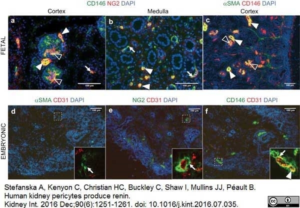
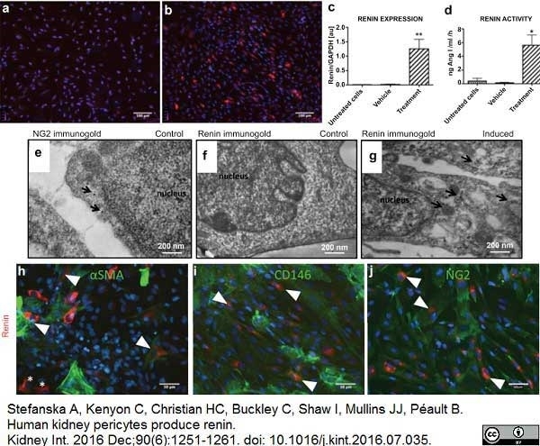
Mouse anti Human CD146 antibody, clone oJ79c (MCA2141) used to identify CD146 expressing pericytes in human fetal and embryonic kidneys by immunofluorescence
Image caption:
Identification of pericytes in human embryonic and fetal kidneys. (a) Fetal pericytes were labeled with antibodies against CD146 (green) and NG2 (red) in a 15-week-old kidney cortex. CD146+NG2+ pericytes were found in peritubular capillaries (arrow), mesangial cells (open arrowheads), and afferent arterioles (arrowheads). (b) In the kidney medulla, pericytes encircled peritubular capillaries (arrows) and the vasa recta (arrowheads). (c) Double staining for CD146 (red) and smooth muscle α-actin (αSMA) (green) shows that in the cortex, pericytes coexpress αSMA in the mesangium (open arrowheads) and afferent arterioles (arrowheads). In embryonic pericytes, sections of 7-week-old kidneys were stained for pericyte markers: CD146 (green), nerve/glial antigen 2 (NG2) (green), and αSMA (green), with the endothelial cell marker CD31 (red) (on consecutive sections). (d) αSMA immunoreactivity was sparse; only single vascular smooth muscle cells were found (inset, arrow). (e) NG2+ cells surrounded endothelial cells (inset, arrow). (f) CD146 staining was perivascular (inset, arrow) and in endothelial cells (inset, arrowhead). DAPI, 4',6-diamidino-2-phenylindole.
From: Stefanska A, Kenyon C, Christian HC, Buckley C, Shaw I, Mullins JJ, Péault B.
Human kidney pericytes produce renin.
Kidney Int. 2016 Dec;90(6):1251-61.
This is from an open access article distributed under the terms of the Creative Commons Attribution License.
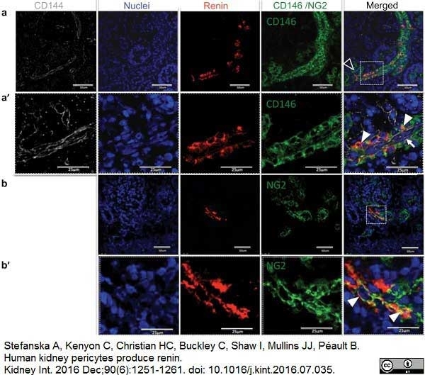
Mouse anti Human CD146 antibody, clone oJ79c (MCA2141) used to identify CD146 expressing pericytes in human fetal kidneys by immunofluorescence.
Image caption:
Renin is colocalized with pericyte markers in the human fetal kidney. Triple staining of fetal kidney demonstrates that (a) renin immunoreactivity (red) coincides with pericyte marker CD146 (green) expression in afferent arterioles. CD146 staining is also present in the mesangium (open arrowhead). Characteristically, renin staining had a striped pattern. The inset (a') shows an afferent arteriole at high magnification. Renin and CD146 stainings overlap on the abluminal side of the vessel (arrowheads), whereas immunoreactivity for CD144 (grey), an endothelial cell marker, is visible inside blood vessels (arrow). (b) Nerve/glial antigen 2 (NG2) (green) staining is found in the mesangium and afferent arteriole. Renin (red) is present in the JG area. Inset (b') shows renin+ cells coexpressing NG2 (arrowheads) at higher magnification. JG, juxtaglomerular cell.
From: Stefanska A, Kenyon C, Christian HC, Buckley C, Shaw I, Mullins JJ, Péault B.
Human kidney pericytes produce renin.
Kidney Int. 2016 Dec;90(6):1251-61.
This is from an open access article distributed under the terms of the Creative Commons Attribution License.
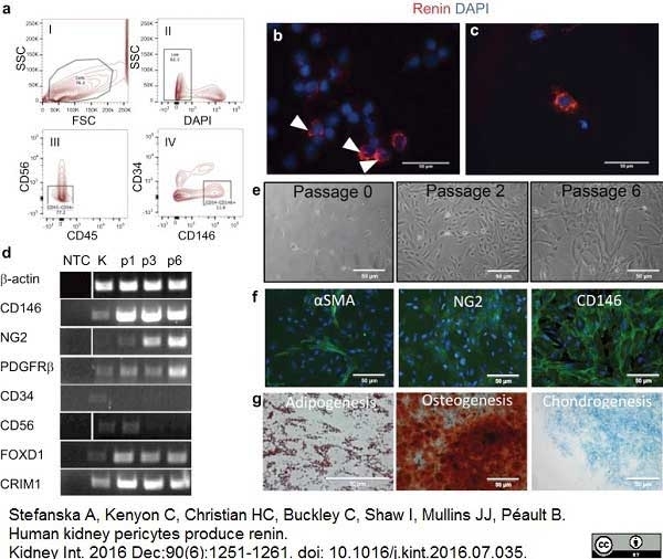
Mouse anti Human CD146 antibody, clone oJ79c (MCA2141) used to identify CD146 expressing pericytes in human fetal kidneys by flow cytometry and to demonstrate CD146 positivity in pericytes cultured in vitro by immunofluorescence.
Image caption:
Isolation and in vitro characterization of pericytes from a human fetal kidney. (a) FACS of renal pericytes purified from a 16-week human fetal kidney. Dot plots show backgating strategy to obtain CD146+CD34−CD56−CD45− pericytes. Red-color dots show gated populations and the percentages of the parent gates. The sorted pericyte population contained no CD45+, CD56+, or CD34+ cells. (b) Cytospins of freshly sorted pericytes were immunostained for renin (red, arrowheads). (c) Granular, intracellular expression of renin (red) can be observed for up to 48 hours in occasional cells of renal pericyte primary cultures. Positively stained cells are native JG cells, which retain renin expression for a short term in vitro. (d) Representative results of gene expression profiles of renal pericyte primary cultures (n ≥ 3). Pericytes showing expression of the pericyte markers CD146, nerve/glial antigen 2 (NG2), and platelet-derived growth factor receptor-β (PDGFRβ), were negative for hematopoietic CD45 and metanephric mesenchyme CD56 expressions. In addition, pericyte cultures were enriched in Foxd1 (stromal marker) and CRIM1 (pericyte/parietal cell/podocyte marker). (e) Morphology of cultured CD146+CD34−CD56−CD45− pericytes. Bright field images of renal pericyte primary cultures are shown after plating at passages (p) 0, 2, and 6. Cells displayed a spindle-shaped morphology at p0 and p2 but eventually acquired fibroblastic-like appearance at p6. (f) Cultured kidney pericytes at p2 show some α-smooth muscle actin (αSMA) (green) positive staining, whereas most of the cells are positive for NG2 (green) and all cultured pericytes are CD146+ (green). (g) Kidney pericyte primary cultures underwent mesodermal lineage induction. After 14 days of differentiation, primary cultures were confirmed to differentiate along adipogenic (Oil red O staining), chondrogenic (Alcian blue staining), and osteogenic lineages (Alizarin red staining). Control cells were incubated with growth medium (not shown). Original magnifications are shown on the images. DAPI, 4',6-diamidino-2-phenylindole; FACS, fluorescence-activated cell sorter; FSC, forward scatter; JG, juxtaglomerular cell; K, kidney (positive control); NTC, no template control; SSC, side scatter.
From: Stefanska A, Kenyon C, Christian HC, Buckley C, Shaw I, Mullins JJ, Péault B.
Human kidney pericytes produce renin.
Kidney Int. 2016 Dec;90(6):1251-61.
This is from an open access article distributed under the terms of the Creative Commons Attribution License.
Mouse anti Human CD146 antibody, clone oJ79c (MCA2141) used to identify CD146 expressing pericytes in human fetal kidneys by immunofluorescence.
Image caption:
CD146+NG2+ αSMA+/− renal pericytes express and secrete enzymatically active renin in vitro. Renal pericyte primary cultures at passage 2 were stained for renin (red). (a) Control cells (vehicle: medium + vehicle; untreated cells: medium) show no renin immunoreactivity. Forskolin and isobutyl-1-methylxanthine (cyclic adenosine monophosphate induction) treatment in pericytes (b) caused robust renin staining, (c) increased renin mRNA expression, and (d) renin activity. One-way analysis of variance followed by Bonferroni's post hoc comparisons were used to test statistical significance. Data are shown as mean ± SEM (n = 3, *P<0.5; n = 5, **P<0.01). Electron micrographs of human pericytes showed immuno-gold labeling for nerve/glial antigen 2 (NG2) (arrows) (e) but (f) no renin immuno-gold labeling. (g) Forskolin-treated pericyte showing immuno-gold labeling for human renin (arrows indicate the immuno-gold label). Renin-induced kidney pericyte primary cultures stained positively for pericyte markers: CD146, NG2, α-smooth muscle actin (αSMA) (all green), and renin (red). (h) Renin expression was not always correlated with αSMA expression (65.68 ± 7.4% overlap). Double positive cells for renin and αSMA are marked with arrowheads and renin+αSMA− cells are indicated by an asterisk. Renin is co-expressed unequivocally (100% overlap) with (i) CD146 and (j) NG2. Double positive cells for renin and pericyte markers are marked with arrowheads. GAPDH, glyceraldehyde-3-phosphate dehydrogenase.
From: Stefanska A, Kenyon C, Christian HC, Buckley C, Shaw I, Mullins JJ, Péault B.
Human kidney pericytes produce renin.
Kidney Int. 2016 Dec;90(6):1251-61.
This is from an open access article distributed under the terms of the Creative Commons Attribution License.
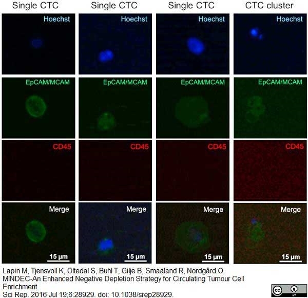
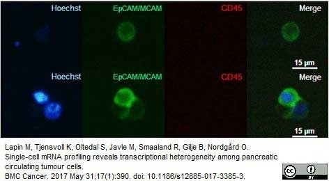
FITC conjugated Mouse anti Human CD146 antibody, clone oJ79c (MCA2141F) used to identify MCAM expression on circulating tumor cells in patients by immunofluorescence.
Image caption:
Representative fluorescence images of CTCs and a CTC cluster isolated from peripheral blood of patients with metastatic pancreatic cancer.
Enriched blood samples were stained with Hoechst 33343 (nuclei, blue), EpCAM (green), MCAM (green), and CD45 (red).
From: Lapin M, Tjensvoll K, Oltedal S, Buhl T, Gilje B, Smaaland R, Nordgård O.
MINDEC-An Enhanced Negative Depletion Strategy for Circulating Tumour Cell Enrichment.
Sci Rep. 2016 Jul 19;6:28929.
This is from an open access article distributed under the terms of the Creative Commons Attribution License.
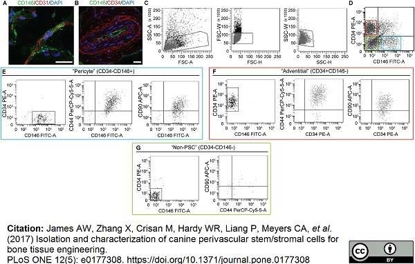
Mouse anti Human CD146 antibody, clone OJ79c (MCA2141) used to identify canine pericytes in situ by immunofluorescence and isolated from adipose tissue by flow cytometry.
Image caption:
Canine PSC in situ identification.
Marker expression and isolation by fluorescence activated cell sorting (FACS). (A) Canine adipose tissue stained with pericytes marker CD146 (green), endothelial marker CD31 (red) (63×), and (B) adventitial cell marker, CD34 (red), CD146 (green) (40×). DAPI nuclear counterstain appears blue. (C) Cell size. (D) Purified PSC consist of distinct CD34−CD146+ pericytes and CD34+CD146− adventitial cells. (E) CD146+CD34- pericytes and (F) CD146-CD34+ adventitial cells express characteristic MSC markers, including CD44 and CD90. In comparison, (G) CD146-CD34- “non-PSC” are predominantly negative for CD44 and CD90. Scale bar: 25μM.
From: James AW, Zhang X, Crisan M, Hardy WR, Liang P, Meyers CA, et al. (2017)
Isolation and characterization of canine perivascular stem/stromal cells for bone tissue engineering.
PLoS ONE 12(5): e0177308.
This is from an open access article distributed under the terms of the Creative Commons Attribution License.
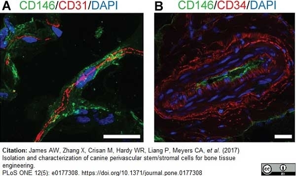
Mouse anti Human CD146 antibody, clone OJ79c (MCA2141) used to identify canine pericytes in situ by immunofluorescence staining of adipose cryosections.
Image caption:
Canine PSC in situ identification.
Marker expression (A) Canine adipose tissue stained with pericytes marker CD146 (green), endothelial marker CD31 (red) (63×), and (B) adventitial cell marker, CD34 (red), CD146 (green) (40×). DAPI nuclear counterstain appears blue. Scale bar: 25μM.
From: James AW, Zhang X, Crisan M, Hardy WR, Liang P, Meyers CA, et al. (2017)
Isolation and characterization of canine perivascular stem/stromal cells for bone tissue engineering.
PLoS ONE 12(5): e0177308.
This is from an open access article distributed under the terms of the Creative Commons Attribution License.
Mouse anti Human CD146 antibody, clone OJ79c (MCA2141) used to identify MCAM expressing circulating tumor cells by immunofluorescence.
Image caption:
Thumbnail gallery of analysed CTCs. Representative fluorescence images of a CTC (top row) and a CTC cluster (bottom row) isolated from the peripheral blood of a patient with pancreatic cancer. (Left column) Hoechst stain (blue) identifies nuclei; (second column) EpCAM-MCAM (green) identifies membranes of CTCs; (third column) CD45 (red) identifies leucocytes; (right column) superimposed images confirms intact cells. Images were acquired with 20× magnification. To enhance visibility, we adjusted the brightness and contrast equally for all microscopic images, and these adjustments were applied to the entire image
From: Lapin M, Tjensvoll K, Oltedal S, Javle M, Smaaland R, Gilje B, Nordgård O.
Single-cell mRNA profiling reveals transcriptional heterogeneity among pancreatic circulating tumour cells.
BMC Cancer. 2017 May 31;17(1):390.
This is from an open access article distributed under the terms of the Creative Commons Attribution License.
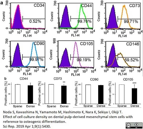
Mouse anti Human CD146 antibody, clone OJ79c (MCA2141) used for the evaluation of CD146 expression on dental pulp derived cells by flow cytometry.
Image caption:
Cell surface markers. (a) Cell surface markers before separation into sparse (sDPSCs) and dense (dDPSCs) groups. A representative case among seven donors is shown. (b) MSC marker expression in sparse (sDPSCs) and dense (dDPSCs) groups. **p = 0.0079 and ***p = 0.0006 (Mann-Whitney U test). The error bar is SD (n = 7). Solid colored histograms represent IgGκ treated cells for control, and open colored histograms represent fluorophore labeled antibody treated cells.
From: Noda S, Kawashima N, Yamamoto M, Hashimoto K, Nara K, Sekiya I, Okiji T.
Effect of cell culture density on dental pulp-derived mesenchymal stem cells with reference to osteogenic differentiation.
Sci Rep. 2019 Apr 1;9(1):5430.
This image is from an open access article distributed under the terms of the Creative Commons Attribution License.
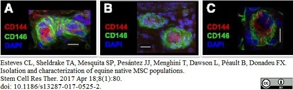
Mouse anti Human CD146 antibody, clone OJ79c (MCA2141) used for the demonstration of equine CD146 expressing cells by immunofluorescence.
Image caption:
Immunohistochemistry of pericyte, adventitial cell, and MSC markers in equine tissues. a–c Pericytes stained for CD146 surrounding CD144+ endothelial cells in adipose tissue (a), testis (b), and skeletal muscle (c). 4′,6-Diamidino-2-phenylindole (DAPI) was used to stain cell nuclei. White scale bars = 10 μm.
From: Esteves CL, Sheldrake TA, Mesquita SP, Pesántez JJ, Menghini T, Dawson L, Péault B, Donadeu FX.
Isolation and characterization of equine native MSC populations.
Stem Cell Res Ther. 2017 Apr 18;8(1):80.
doi: 10.1186/s13287-017-0525-2.
This image is from an open access article distributed under the terms of the Creative Commons Attribution License.
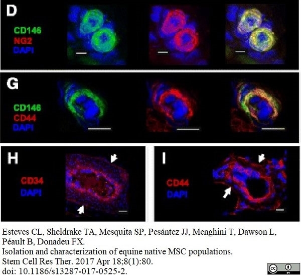
Mouse anti Human CD146 antibody, clone OJ79c (MCA2141) used for the demonstration of equine CD146 expressing cells by immunofluorescence.
Image caption:
Immunohistochemistry of pericyte, adventitial cell, and MSC markers in equine tissues. d, Dual staining (right panel) with the pericyte markers NG2 and CD146 (d) in adipose tissue. g, Colocalization (right panel) of the MSC and pericyte markers CD44 and CD146 (g) in adipose tissue. d–g Individual staining is shown (left and middle panels). h, i Immunodetection of the adventitial cell marker CD34 (h) and the MSC marker CD44 (i) in the outer layer of blood vessels (arrows) in adipose tissue. 4′,6-Diamidino-2-phenylindole (DAPI) was used to stain cell nuclei. White scale bars = 10 μm.
From: Esteves CL, Sheldrake TA, Mesquita SP, Pesántez JJ, Menghini T, Dawson L, Péault B, Donadeu FX.
Isolation and characterization of equine native MSC populations.
Stem Cell Res Ther. 2017 Apr 18;8(1):80.
doi: 10.1186/s13287-017-0525-2.
This image is from an open access article distributed under the terms of the Creative Commons Attribution License.
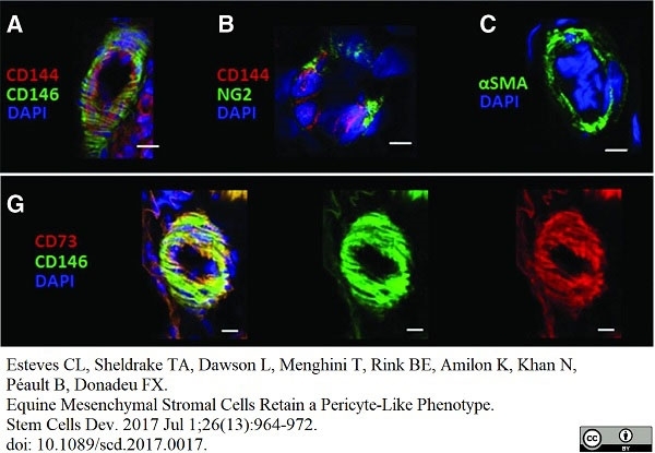
FITC conjugated Mouse anti CD146 antibody, clone OJ79c (MCA2141F) used to demonstrate CD146 expressing equine pericytes by immunofluorescence.
Image caption:
IHC of equine AT sections showing staining for the pericyte markers, CD146 (A), NG2 (B), αSMA (C),(red; A, B ). Colocalization of MSC markers CD146 and CD73; F, G, in yellow (left panel) from the overlap of green (middle panel) and red (right panel) individual antibody fluorescence. DAPI was used to stain nuclei. Scale bar, 10 &mu,m, is indicated by white bars. IHC, immunohistochemistry; MSC, mesenchymal stem/stromal cell. l.
From: Esteves CL, Sheldrake TA, Dawson L, Menghini T, Rink BE, Amilon K, Khan N, Péault B, Donadeu FX.
Equine Mesenchymal Stromal Cells Retain a Pericyte-Like Phenotype.
Stem Cells Dev. 2017 Jul 1;26(13):964-72.
doi: 10.1089/scd.2017.0017.
This image is from an open access article distributed under the terms of the Creative Commons Attribution License.
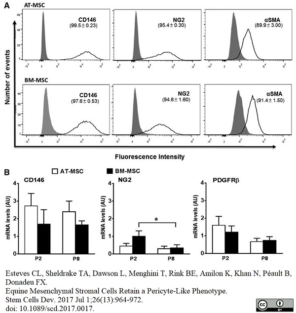
Alexa-Fluor®647 conjugated Mouse anti Human CD146 antibody, clone OJ79c (MCA2141A647) used to assess CD146 nexpression on adipose tissue-derived and mone marrow-derived mesenchymal stem cells by flow cytometry.
Image caption:
(A) Flow cytometry histograms showing the proportions of AT-MSCs and BM-MSCs (upper and lower panels, respectively) that are positive for staining with the antibodies CD146-AF647, NG2-APC, and αSMA (with secondary AF405 conjugated). Isotype controls are shown by the gray peak and unstained curves are omitted for simplicity. (B) Results of qPCR analysis of CD146, NG2, and PDGFRβ in AT-MSCs and BM-MSCs at passages 2 (P2) and 8 (P8). All results are shown as mean ± SEM; n = 3 animals. *P <0.05.
From: Esteves CL, Sheldrake TA, Dawson L, Menghini T, Rink BE, Amilon K, Khan N, Péault B, Donadeu FX.
Equine Mesenchymal Stromal Cells Retain a Pericyte-Like Phenotype.
Stem Cells Dev. 2017 Jul 1;26(13):964-72.
doi: 10.1089/scd.2017.0017.
This image is from an open access article distributed under the terms of the Creative Commons Attribution License.
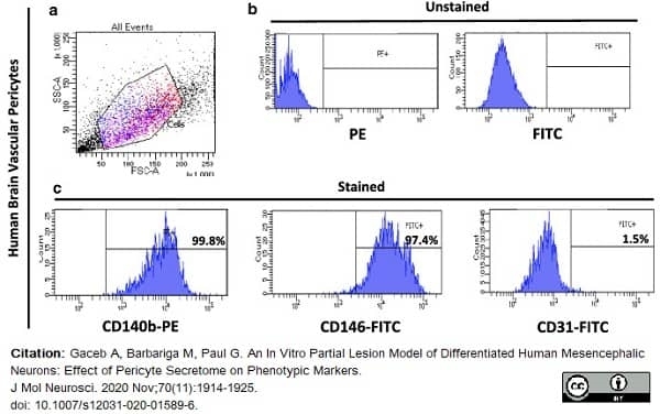
FITC conjugated Mouse anti Human CD146 antibody, clone OJ79c (MCA2141F) used to evaluate CD146 expression on human brain vascular pericytes by flow cytometry.
Image caption:
Characterisation of human brain vascular pericytes by flow cytometry. (A) Cells were identified according to forward scatter (FSC) and side scatter (SSC) intensity. (B) Histograms of unstained human brain pericytes. (C) Histograms represent the fluorescence intensity of human brain pericytes after incubation with CD140b, CD146, and CD31 antibodies, respectively. (3 independent experiments, 3 replicates)
From: Gaceb A, Barbariga M, Paul G.
An In Vitro Partial Lesion Model of Differentiated Human Mesencephalic Neurons: Effect of Pericyte Secretome on Phenotypic Markers.
J Mol Neurosci. 2020 Nov;70(11):1914-1925.
doi: 10.1007/s12031-020-01589-6.
This image is from an open access article distributed under terms of a Creative Commons Attribution License.
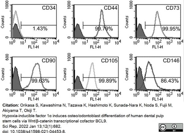
Mouse anti Human CD146 antibody, clone OJ79c (MCA2141) used to assess CD146 expression on cultured mesenchymal stem cells by flow cytometry.
Image caption:
hDPSCs express typical mesenchymal stem cell (MSC) markers and exhibit multi-differentiation potential. (a) Typical MSC markers (CD44, CD73, CD90, CD105, CD146) are highly expressed in hDPSCs, and hDPSCs are mostly negative for a hematopoietic marker (CD34). hDPSCs possess neurogenic, adipogenic, chondrogenic, and osteogenic differentiation potential, as determined by the expression of neurogenic markers (GFAP and NF-M)
From: Orikasa S, Kawashima N, Tazawa K, Hashimoto K, Sunada-Nara K, Noda S, Fujii M, Akiyama T, Okiji T.
Hypoxia-inducible factor 1α induces osteo/odontoblast differentiation of human dental pulp stem cells via Wnt/β-catenin transcriptional cofactor BCL9.
Sci Rep. 2022 Jan 13;12(1):682.
doi: 10.1038/s41598-021-04453-8.
This image is from an open access article distributed under terms of a Creative Commons Attribution License.
Mouse anti Human CD146
- Product Type
- Monoclonal Antibody
- Clone
- OJ79c
- Isotype
- IgG1
- Specificity
- CD146
| Mouse anti Human CD146 antibody, clone OJ79c recognizes human Cell surface glycoprotein MUC18, also known as CD146, Cell surface glycoprotein P1H12, Melanoma cell adhesion molecule (MCAM) or S-endo 1 endothelial-associated antigen. CD146 is a 646 amino acid single pass type 1 transmembrane glycoprotein with a calculated molecular mass of ~72 kDa. However due to extensive N-linked glycosylation CD146 migrates in polyacrylamide gels with an apparent molecular mass of ~118 kDa. CD146 is a member of the immunoglobulin superfamily bearing 2 V-type Ig-like and 3 C-type Ig-like domains. CD146 is expressed by all endothelial cells and by melanoma cells and appears to act as an adhesion molecule (UniProt: P43121). Expression in melanoma may be linked to tumor progression (Lehmann et al. 1989). Mouse anti Human CD146 antibody, clone OJ79c is highly expressed on pericytes and has been utilized for the identification of perivascular mesenchymal precursor cells from cardiac muscle using flow cytometry (Chen et al. 2014). |
- Target Species
- Human
- Species Cross-Reactivity
-
Target Species Cross Reactivity Pig Dog - N.B. Antibody reactivity and working conditions may vary between species.
- Product Form
- Purified IgG - liquid
- Preparation
- Purified IgG prepared by affinity chromatography on Protein A from tissue culture supernatant
- Buffer Solution
- Phosphate buffered saline
- Preservative Stabilisers
- 0.09% sodium azide (NaN3)
- Carrier Free
- Yes
- Immunogen
- Recombinant human MUC18 (D1-D5) Fc protein.
- Approx. Protein Concentrations
- IgG concentration 1.0 mg/ml
- Fusion Partners
- Spleen cells from immunized mice were fused with cells of the mouse Sp2/0 Ag.14 myeloma cell line.
- Regulatory
- For research purposes only
- Guarantee
- 12 months from date of despatch
This product is shipped at ambient temperature. It is recommended to aliquot and store at -20°C on receipt. When thawed, aliquot the sample as needed. Keep aliquots at 2-8°C for short term use (up to 4 weeks) and store the remaining aliquots at -20°C.
Avoid repeated freezing and thawing as this may denature the antibody. Storage in frost-free freezers is not recommended.
Avoid repeated freezing and thawing as this may denature the antibody. Storage in frost-free freezers is not recommended.
This product has been reported to work in the following applications. This information is derived from testing within our laboratories, peer-reviewed publications or personal communications from the originators. Please refer to references indicated for further information. For general protocol recommendations, please visit the antibody protocols page.
| Application Name | Verified | Min Dilution | Max Dilution |
|---|---|---|---|
| ELISA | |||
| Flow Cytometry | 1/10 | 1/50 | |
| Immunofluorescence | |||
| Immunohistology - Frozen |
Where this product has not been tested for use in a particular technique this does not necessarily exclude its use in such procedures. Suggested working dilutions are given as a guide only. It is recommended that the user titrates the product for use in their own system using appropriate negative/positive controls.
- Flow Cytometry
- Use 10μl of the suggested working dilution to label 106 cells in 100μl
- Histology Positive Control Tissue
- Melanoma
| Description | Product Code | Applications | Pack Size | List Price | Your Price | Quantity | |
|---|---|---|---|---|---|---|---|
| Mouse IgG1 Negative Control | MCA928 | F | 100 Tests |

|
Log in | ||
| List Price | Your Price | ||||||
|
|
Log in | ||||||
| Description | Mouse IgG1 Negative Control | ||||||
References for CD146 antibody
-
Paul, G. et al. (2012) The adult human brain harbors multipotent perivascular mesenchymal stem cells.
PLoS One. 7: e35577. -
Crisan, M. et al. (2008) A perivascular origin for mesenchymal stem cells in multiple human organs.
Cell Stem Cell. 3: 301-13. -
Iohara, K. et al. (2008) A novel stem cell source for vasculogenesis in ischemia: subfraction of side population cells from dental pulp.
Stem Cells. 26 (9): 2408-18. -
Park, T.S. et al. (2010) Placental Perivascular Cells for Human Muscle Regeneration.
Stem Cells Dev. 20: 451-63. -
Smith, K. et al. (2011) Mono- and tri-cationic porphyrin-monoclonal antibody conjugates: photodynamic activity and mechanism of action.
Immunology. 132: 256-65. -
James, A.W. et al. (2012) Perivascular stem cells: a prospectively purified mesenchymal stem cell population for bone tissue engineering.
Stem Cells Transl Med. 1 (6): 510-9. -
Wassmer, S.C. et al. (2011) Vascular endothelial cells cultured from patients with cerebral or uncomplicated malaria exhibit differential reactivity to TNF.
Cell Microbiol. 13: 198-209. -
Lee, J.H. et al. (2012) Generation of osteogenic construct using periosteal-derived osteoblasts and polydioxanone/pluronic F127 scaffold with periosteal-derived CD146 positive endothelial-like cells.
J Biomed Mater Res A.101: 942-53.
View The Latest Product References
-
Boneberg, E.M. et al. (2009) Soluble CD146 is generated by ectodomain shedding of membrane CD146 in a calcium-induced, matrix metalloprotease-dependent process.
Microvasc Res. 78: 325-31. -
Nielsen, C.T. et al. (2011) Distinct features of circulating microparticles and their relationship to clinical manifestations in systemic lupus erythematosus.
Arthritis Rheum. 63: 3067-77. -
Iversen, L.V. et al. (2013) A heparin-based method for flow cytometric analysis of microparticles directly from platelet-poor plasma in calcium containing buffer.
J Immunol Methods. 388 (1-2): 49-59. -
Meireles, A.L. et al. (2011) Increased levels of circulating endothelial progenitor cells in human T-cell lymphotropic virus type I carriers.
Arch Med Res. 42: 34-7. -
Iversen, L.V. et al. (2013) Circulating microparticles and plasma levels of soluble E- and P-selectins in patients with systemic sclerosis.
Scand J Rheumatol. 42 (6): 473-82. -
Murakami, M. et al. (2013) The use of granulocyte-colony stimulating factor induced mobilization for isolation of dental pulp stem cells with high regenerative potential.
Biomaterials. pii: S0142-9612(13)00942-3. -
Ruetze, M. et al. (2013) A novel niche for skin derived precursors in non-follicular skin.
J Dermatol Sci. 69: 132-9. -
Dokić, J. et al. (2013) Mesenchymal stem cells from periapical lesions modulate differentiation and functional properties of monocyte-derived dendritic cells.
Eur J Immunol. 43: 1862-72. -
Chen, W.C. et al. (2015) Human myocardial pericytes: multipotent mesodermal precursors exhibiting cardiac specificity.
Stem Cells. 33 (2): 557-73. -
Iversen, L.V. et al. (2015) Cross-sectional study of soluble selectins, fractions of circulating microparticles and their relationship to lung and skin involvement in systemic sclerosis.
BMC Musculoskelet Disord. 16: 191. -
Tawonsawatruk T et al. (2016) Adipose derived pericytes rescue fractures from a failure of healing - non-union.
Sci Rep. 6: 22779. -
Boissier, R. et al. (2016) Histologic and urodynamic effects of autologous stromal vascular fraction extracted from fat tissue with minimal ex vivo manipulation on a porcine model of intrinsic sphincter deficiency
J Urology. Jun 2 [Epub ahead of print] -
Esteves, C.L. et al. (2017) Equine Mesenchymal Stromal Cells Retain a Pericyte-Like Phenotype.
Stem Cells Dev. 26 (13): 964-72. -
Stefanska, A. et al. (2016) Human kidney pericytes produce renin.
Kidney Int. 90 (6): 1251-61. -
Lapin, M. et al. (2016) MINDEC-An Enhanced Negative Depletion Strategy for Circulating Tumour Cell Enrichment.
Sci Rep. 6: 28929. -
Muerza-Cascante, M.L. et al. (2016) Endosteal-like extracellular matrix expression on melt electrospun written scaffolds.
Acta Biomater. pii: S1742-7061(16)30706-1. -
Eliasberg, C.D. et al. (2017) Perivascular Stem Cells Diminish Muscle Atrophy Following Massive Rotator Cuff Tears in a Small Animal Model.
J Bone Joint Surg Am. 99 (4): 331-41. -
James, A.W. et al. (2017) Isolation and characterization of canine perivascular stem/stromal cells for bone tissue engineering.
PLoS One. 12 (5): e0177308. -
Shen, J. et al. (2018) Effects of WNT3A and WNT16 on the Osteogenic and Adipogenic Differentiation of Perivascular Stem/Stromal Cells.
Tissue Eng Part A. 24 (1-2): 68-80. -
Lapin, M. et al. (2017) Single-cell mRNA profiling reveals transcriptional heterogeneity among pancreatic circulating tumour cells.
BMC Cancer. 17 (1): 390. -
Esteves, C.L. et al. (2017) Isolation and characterization of equine native MSC populations.
Stem Cell Res Ther. 8 (1): 80. -
Gaceb, A. et al. (2017) Pericytes secrete pro-regenerative molecules in response to platelet-derived growth factor-BB.
J Cereb Blood Flow Metab. : 271678X17719645. -
Noda, S. et al. (2019) Effect of cell culture density on dental pulp-derived mesenchymal stem cells with reference to osteogenic differentiation.
Sci Rep. 9 (1): 5430. -
Gaceb, A. et al. (2020) An In Vitro. Partial Lesion Model of Differentiated Human Mesencephalic Neurons: Effect of Pericyte Secretome on Phenotypic Markers.
J Mol Neurosci. 70 (11): 1914-25. -
Stefanska, A. et al. (2021) Role of Pericytes in the Development of the Renin/Angiotensin System: Induction of Functional Renin in Cultures of Pericytes.
Methods Mol Biol. 2235: 169-180. -
Orikasa, S. et al. (2022) Hypoxia-inducible factor 1α induces osteo/odontoblast differentiation of human dental pulp stem cells via Wnt/β-catenin transcriptional cofactor BCL9.
Sci Rep. 12 (1): 682. -
Menon, R. et al. (2023) Human Induced Pluripotent Stem Cell-Derived Pericytes as Scalable and Editable Source to Study Direct Lineage Reprogramming Into Induced Neurons.
Cell Reprogram. 25 (5): 212-23. -
Molinos, M. et al. (2023) Alterations of bovine nucleus pulposus cells with aging.
Aging Cell. 22 (8): e13873. -
An, H.J. et al. (2021) Pro-Angiogenic and Osteogenic Effects of Adipose Tissue-Derived Pericytes Synergistically Enhanced by Nel-like Protein-1.
Cells. 10 (9): 2244.
Further Reading
-
Kuzu, I. et al. (1993) Expression of adhesion molecules on the endothelium of normal tissue vessels and vascular tumors.
Lab Invest. 69 (3): 322-8. -
Piriou-Guzylack, L. (2008) Membrane markers of the immune cells in swine: an update.
Vet Res. 39: 54.
- Synonyms
- MUC18
- RRID
- AB_324068
- UniProt
- P43121
- Entrez Gene
- MCAM
- GO Terms
- GO:0005886 plasma membrane
- GO:0007155 cell adhesion
- GO:0016021 integral to membrane
- GO:0009653 anatomical structure morphogenesis
Request a different product with this specificity
Please Note: All Products are "FOR RESEARCH PURPOSES ONLY"
View all Anti-Human ProductsAlways be the first to know.
When we launch new products and resources to help you achieve more in the lab.
Yes, sign me up