Popular topics

-
References
Austyn JM and Gordon S. (1981). F4/80, a monoclonal antibody directed specifically against the mouse macrophage. Eur J Immunol 11, 805-81
Gordon S and Plüddemann A. (2017). Tissue macrophages: heterogeneity and functions. BMC Biol 15, 53
Hume DA et al. (1984). The mononuclear phagocyte system of the mouse defined by immunohistochemical localisation of antigen F4/80: Macrophages associated with epithelia. Anat Rec 210, 503-512
Lin HH et al. (2005). The macrophage F4/80 receptor is required for the induction of antigen-specific efferent regulatory T cells in peripheral tolerance. J Exp Med 201, 1615-1625
McKnight AJ and Gordon S. (1998). The EGF-TM7 family: unusual structures at the leukocyte surface. J Leukoc Biol 63, 271-280
Morris L et al. (1991). Macrophages in haemopoietic and other tissues of the developing mouse detected by the monoclonal antibody F4/80. Development 112, 517-526
Perry VH et al. (1985). Immunohistochemical localization of macrophages and microglia in the adult and developing mouse brain. Neuroscience 15, 313-326
Siamon Gordon: The Father of F4/80
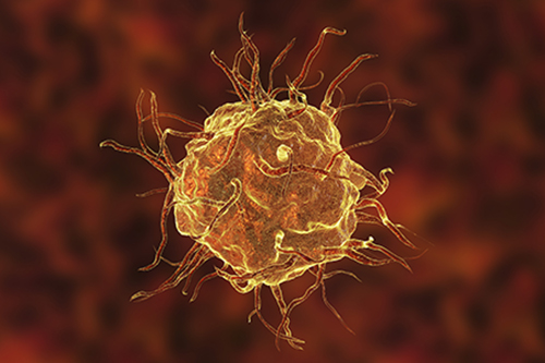
F4/80, a member of the epidermal growth factor-seven transmembrane (EGF-TM7) family, is arguably the best-known mouse macrophage and microglia marker. The clone Cl:A3-1 is well-characterized and extensively referenced, with an established heritage going back to 1981 (Austyn and Gordon 1981). Cl:A3-1 was isolated by Siamon Gordon's group over 40 years ago. We spoke to Professor Gordon about his career in immunology and love of macrophages.
From growing up in a small village in South Africa and learning English at 15, to an impressive scientific pedigree and contributions to macrophage research over the past 50 years, in this blog Siamon Gordon tells us about his life and his research.

Professor Siamon Gordon
Bio-Rad (BR): Could you tell us a bit about how you chose a career in research?
Siamon Gordon (SG): I grew up in Darling, a small village near Cape Town, South Africa, and didn’t learn English until I was 15, when I moved to an English medium school. If you grew up in my Jewish world, there weren’t many options when it came to career choices. It was expected that you would be a doctor, lawyer, pharmacist, or an accountant. My mother thought that I would hate the sight of blood, but I decided medicine was the option for me, as it opens your mind to a completely different world. So, I attended the University of Cape Town to study medicine. I started with the basic sciences, botany, and zoology, then later studied anatomy and physiology, before studying pathology. It wasn’t until in my fourth year that I became exposed to clinical medicine, surgery, and then obstetrics, gynecology, and so on. I worked in a hospital called Groote Schuur Hospital, where I interned with professors of medicine and surgery, including a stint at Red Cross Children's Hospital.
I was always interested in my subject, and I would want to know why something happened, and I decided to go into research to understand the “why”. At the time, I was waiting for my then-girlfriend, who later became my wife, to finish her undergraduate course in English literature at the University of Cape Town, so I did an extra year in the labs doing basic diagnostics, like histopathology, microbiology, and clinical chemistry. That was the last time I had any real contact with medicine.
BR: How did you decide where to go next?
SG: In the early 1960s, whenever one graduated in medicine you needed to acquire specialist training, and this meant leaving South Africa. I left South Africa, and like many other emigrants, never went back there to live, but I’ve always tried to give back, as I owe a lot to the country to which my parents had immigrated from Eastern Europe. When I was planning where to go for my research experience, I got advice from a very supportive mentor, Dr Golda Selzer, who put me in contact with someone in London and someone in New York. The person in New York was slow to respond, so I accepted the English offer and then when the other position came through as well, I made them consecutive rather than having to choose only one. I first went to the Wright-Fleming Institute at St. Mary’s Hospital in London, to the lab of Rodney Porter, who was working on antibody structure and separation of the immunoglobulin chains. That was the first real immunology I encountered. After a year in London, I went to Rockefeller University in New York to a human genetics lab run by Alexander Bearn. I spent a year with him but knew already that I needed much more research experience and science education, so I decided to go back to being a graduate student. I spent the next five years, from 1966 to 1971, completing my doctoral degree and stayed on at Rockefeller until 1976.
BR: How did you become interested in macrophages?
SG: Well, I was extremely lucky because when I was looking for a project, I knew I didn’t want to work on proteins only, and I thought I'd rather work on cell biology. By chance, Henry Harris, who was at the Sir William Dunn School of Pathology in Oxford, was visiting after he had published his wonderful paper on using irradiated Sendai virus to fuse cells from different species or different cell types. It seemed like a really exciting opportunity and I thought that's what I'd like to do. I needed to find a host laboratory that would be interested in this type of project. Thankfully, the lab of Zanvil Cohn and James Hirsch was open to this idea. Hirsch was a pioneer in neutrophil research and Cohn was interested in macrophages. A thing at the time that made it particularly interesting is that unlike other labs, we used phase contrast microscopy to look at the live cultures. That's when I got hooked on macrophages because they were very aesthetically pleasing, they ruffle and I could see the wonderful cytoplasmic details with all the organelles. That was it, from then on, I worked on macrophages.
BR: How did you come to discover F4/80?
SG: In 1976, we moved to Oxford as a family on a long-term basis and I started my own lab for the first time. Around that time, monoclonal antibody technology had appeared on the horizon. We knew already that if you fuse cells that were different, you actually extinguished some of their specialized properties and you picked up things like immortality. Using this method, Milstein and Kohler were able to clone antibody-producing B cells via fusion with myeloma cells, allowing the production of antigen-specific monoclonal antibodies. They later won a Nobel Prize in Physiology or Medicine for this technology. Since I had a background in cell fusion, my first goal was to make a monoclonal antibody that would be specific for macrophages. My first graduate student, Jon Austyn, fused mouse macrophages with rat myeloma cells and screened the antibodies on primary mouse macrophages. We were lucky that the first specific antibody we produced was the F4/80 one that is still available today. The antibodies were screened on thioglycolate-elicited peritoneal macrophages after glutaraldehyde fixation. This was a fortunate choice because this harsh protocol meant that the F4/80 antigen could be studied in tissue sections. We were also fortunate that the antigen had not been described before and was a plasma membrane marker, which was very useful for immunohistochemistry (Figure 1). Macrophages stain beautifully with this antibody on paraformaldehyde-fixed sections; one can readily see their cell processes and interactions with neighboring cells. Other markers don't do this as well, which is the beauty of the F4/80.

Fig 1. Mouse spleen cryosection stained with Rat anti-Mouse F4/80 antibody, clone Cl:A3-1 (brown). Note staining of red, but not white pulp.
BR: What would you say was the impact of discovering F4/80?
SG: This was the start of macrophage phenotypic characterization. You could look anywhere, for the first time, and would see the extent of the dispersed mononuclear phagocyte system, which defines monocytes, macrophages, possibly some dendritic cells, and also giant cells (Hume et al. 1984). Although I should say there are one or two interesting cells that are macrophages for sure, but don't have F4/80, such as metallophilic cells in the splenic marginal zone; so we screened again on such tissues to see if we could find another antibody that would mark those cells. That's how we identified other macrophage markers, such as Siglec-1 (CD169), a sialic acid-dependent antigen. Other lectin-like receptors studied include the macrophage mannose receptor (CD206) and Dectin-1 and unrelated scavenger receptors. Both the F4/80 and the Siglec-1 antigens became founders of distinct macrophage-restricted glycoprotein families.
BR: Why do you think it’s important to understand macrophages?
SG: Macrophages recognize things that are changed physiologically and help to restore them if possible. One of the famous quotes that I was exposed to from the geneticist Theodosius Dobzhansky when I was a student, was “nothing in biology makes sense except in the light of evolution”. That’s lesson number one. I have a different version, relevant to macrophages, which is “nothing in macrophage biology makes sense except in the light of homeostasis”. It’s much more than immunology; development, physiology, and many disease processes have at their root the need for a cell like the macrophage to be present everywhere and able to perform a variety of specialized functions (Gordon and Plüddermann 2017, Lin et al 2005, Morris et al. 1991, Perry et al. 1985). Their main housekeeping function is to get rid of dying cells and worn-out molecules, breaking down and recycling molecules. The “self” that exists now isn't made of the same molecules as the ones that you start with. You keep rebuilding the body. You need macrophages to be able to do this in trophic as well as digestive functions and to interact with other cell types in the body.
BR: What advice would you give to early career scientists?
SG: To expect a lot of disappointments, to dream of science, to have imagination, and to put yourself in the position of a germ to understand what their strategy needs to be versus yours. I think the most important thing is to keep a sense of wonder. Biology is fantastically interesting, and marvelous. You just have to say, “It’s a miracle, how does this happen?”.
BR: You mentioned before that you try to give back to South Africa. Can you tell us more about this?
SG: Yes, South Africa had one of the worst HIV epidemics, and still has more people living with HIV than most other countries. We weren’t working on HIV. We weren’t working on vaccination against HIV. We didn’t have antiretroviral drugs. We weren’t interested in drug development, so decided to produce a cartoon booklet called “You, Me and HIV” aimed at health workers and teachers, educating adolescent children, a form of social vaccination, to know your status and not spread infection to other people, etc. We published it in English, Afrikaans, and Zulu and distributed about 90,000 copies to schools, and employed a social worker who met with teachers and did school visits to raise awareness.
Thank you Professor Siamon Gordon for sharing your inspiring journey with us. We hope that our readers will be motivated by your story and advice to pursue their own great scientific achievements.
Want to Try the F4/80 Antibody for Yourself?
Bio-Rad is the only commercial manufacturer of anti-F4/80 clone CI:A3-1, the original monoclonal antibody produced against the F4/80 antigen in Siamon Gordon’s laboratory.
References
Austyn JM and Gordon S. (1981). F4/80, a monoclonal antibody directed specifically against the mouse macrophage. Eur J Immunol 11, 805-81
Gordon S and Plüddemann A. (2017). Tissue macrophages: heterogeneity and functions. BMC Biol 15, 53
Hume DA et al. (1984). The mononuclear phagocyte system of the mouse defined by immunohistochemical localisation of antigen F4/80: Macrophages associated with epithelia. Anat Rec 210, 503-512
Lin HH et al. (2005). The macrophage F4/80 receptor is required for the induction of antigen-specific efferent regulatory T cells in peripheral tolerance. J Exp Med 201, 1615-1625
McKnight AJ and Gordon S. (1998). The EGF-TM7 family: unusual structures at the leukocyte surface. J Leukoc Biol 63, 271-280
Morris L et al. (1991). Macrophages in haemopoietic and other tissues of the developing mouse detected by the monoclonal antibody F4/80. Development 112, 517-526
Perry VH et al. (1985). Immunohistochemical localization of macrophages and microglia in the adult and developing mouse brain. Neuroscience 15, 313-326
You may also be interested in...

View more Careers or Immunology blogs
