Popular topics

Popular topics
-
References
Bustos R and Sobrino F (1992). Stimulation of glycolysis as an activation signal in rat peritoneal macrophages. Effect of glucocorticoids on this process. Biochem J 282, 299-303.
Hooftman A et al. (2023). Macrophage fumarate hydratase restrains mtRNA-mediated interferon production. Nature 615, 490-498.
Hou F et al. (2011). MAVS forms functional prion-like aggregates to activate and propagate antiviral innate immune response. Cell 146, 448-461.
Wang F et al. (2018). Glycolytic stimulation is not a requirement for M2 macrophage differentiation. Cell Metab 28, 463-475.
Zecchini V et al. (2023). Fumarate induces vesicular release of mtDNA to drive innate immunity. Nature 615, 499-506.
From Powerhouses to Inferno: A Mitochondrial Messenger Fuels Inflammation
The crosstalk between metabolism and immunity, also known as immunometabolism, is an emerging paradigm in understanding immune activation. Previous studies have indicated that metabolic changes are critical to support the functions of certain immune cells. A classic example is metabolic rewiring in macrophage polarization, a process by which macrophages are differentiated into different states based on environmental cues. For instance, when macrophages are differentiated into the M1 lineage, characterized by pro-inflammatory signatures, the cells will enhance the rate of glycolysis (Bustos and Sobrino 1992). In contrast, inhibition of glycolysis will promote anti-inflammatory properties in the M2 lineage (Wang et al. 2018).
While the role of metabolic reprogramming in immune cells is well-documented, the precise mechanism by which metabolism can directly shape immune responses remains unclear.
In this blog, we discuss the concept of immunometabolism, including a new study published by Hooftman et al. (2023) in Nature showing how a molecule derived from mitochondrial metabolism can trigger inflammation, providing insights into the versatile functions of these organelles beyond energy production.
Mitochondria Are Signaling Hubs for Immunity
Mitochondria play pivotal roles in the regulation of various cellular processes, particularly in energy production. These dynamic organelles harbor their own nucleic acids called mitochondrial DNA (mtDNA), which are transcribed into mitochondrial RNA (mtRNA). When mitochondria are impaired, mitochondrial stress can occur, leading to the release of both mtDNA and mtRNA into the cytosol. The release of mitochondrial nucleic acids can subsequently trigger an immune response.
Now, how does the release of mitochondrial nucleic acid activate an immune response?
It appears that mitochondria are not only recognized as the primary energy source for cells but also function as a platform for immune signaling. Foreign nucleic acids such as cytosolic mtRNA or viral RNA can be detected by nucleic acid sensors including toll-like receptor 7(TLR-7), retinoic acid-inducible gene I (RIG-I), and melanoma differentiation-associated protein 5 (MDA5). Specifically, RIG-I or MDA5 can detect these aberrant RNAs and activate a mitochondrial protein called mitochondrial antiviral signaling (MAVS). Activation of MAVS induces the expression of interferon-ß (IFN-ß), an inflammatory cytokine that initiates an antiviral response (Hou et al. 2011).
A Buildup of a Metabolite in Mitochondria Drives Inflammation
A research group from Trinity Biomedical Sciences Institute in Dublin found that the accumulation of a metabolic byproduct from mitochondria can overwhelm the immune system (Hooftman et al. 2023).
To mine for the metabolite associated with inflammation, the researchers used a tool called liquid chromatography-mass spectrometry (LC-MS) to measure changes in molecules inside the cells. Notably, the scientists identified fumarate as one of the most abundant molecules in mouse macrophages treated with lipopolysaccharide (LPS), a pro-inflammatory stimulus.
Fumarate is a metabolite derived from the tricarboxylic acid (TCA) cycle, a metabolic pathway that takes place in the mitochondria and generates energy by breaking down a molecule called acetyl coenzyme A (acetyl-CoA) into carbon dioxide and water. Inside mitochondria, fumarate can be converted into a different metabolic compound named malate by fumarate hydratase (FH). Both genetic deletion (Fh1-/-) and pharmacological inhibition of FH with a drug (FHIN1) resulted in elevated levels of fumarate in LPS-stimulated macrophages. By leveraging both methods, the research group found either loss or suppression of FH led to mitochondrial stress and dysregulation of inflammatory cytokine expression.
So, the key question is: how does the buildup of fumarate drive inflammation?
Using RNA-seq analysis, the study group discovered many IFN-stimulated genes (ISGs) are upregulated in LPS-primed macrophages treated with FHIN1 compared to DMSO treated controls. As mitochondrial stress was induced in FHIN1-treated cells, the researchers proposed the buildup of fumarate can trigger the release of mitochondrial nucleic acids, which can be detected by immune sensors to initiate the IFN response (Fig. 1). Consistently, LPS-primed Fh1-/- macrophages show increased IFN-ß levels, and FHIN1 treatment in cells led to the release of mtRNA in the cytosol. Moreover, induction of IFN-ß depends on MDA5/RIG-I/TLR7, as genetic silencing of these sensors does not change IFN-ß expression in the presence of LPS/FHIN1. Notably, in vivo experiments also show mice administered with LPS and FHIN1 displayed enhanced IFN-ß response.

Fig. 1. The accumulation of fumarate in mitochondria will activate the IFN response. Suppression of fumarate hydrase with the inhibitor, FHIN1, triggers the release of mtRNA and IFN-ß expression.
Low Expression and Defects in Fumarate Hydrase Are Implicated in Human Diseases
Dysregulation in the IFN response drives certain diseases like systemic lupus erythematosus (SLE), an autoimmune condition marked by widespread inflammation. Prior reports have demonstrated mitochondrial dysfunction and induction of mitochondrial stress in blood samples obtained from individuals diagnosed with SLE. Strikingly, Hooftman et al. also found reduced expression of FH in whole blood from SLE patients, indicating that FH suppression may potentially serve as an underlying mechanism for the development of SLE.
Interestingly, another study by Zecchini et al. (2023) in Nature also reports similar findings. This separate group discovered elevated ISG response in kidney cells from Fh1-/- mice, and also found increased fumarate levels in murine kidney cells without FH using LC-MS. This research demonstrated that both the loss of FH and treatment with a derivative of fumarate, monomethyl fumarate (MMF), led to the release of mtDNA into the cytosol where it is detected by a DNA sensor called cyclic GMP–AMP synthase (cGAS) to trigger the IFN response. Additionally, the study group observed a higher abundance of mitochondrial-derived vesicles (MDV) in cells lacking FH under the microscope, suggesting their potential involvement in the release of mtDNA. Indeed, genetic silencing of SNX9, a budding protein required for the formation of MDV, also reduced the release of mtDNA in cells treated with MMF. The loss of FH often occurs in a type of kidney cancer called hereditary leiomyomatosis and renal cell carcinoma (HLRCC) therefore, the study group examined gene expression of tumor tissues from HLRCC patients to look at the inflammatory response. They found an increased inflammatory response in HLRCC tissues, implying that chronic inflammation might be a key driving factor in HLRCC. Overall, this independent study identified a potential link between mtDNA release and HLRCC.
Collectively, these studies highlight the role of mitochondrial metabolism in inflammation and provide a new perspective for the development of therapeutic strategies to treat these inflammatory diseases.
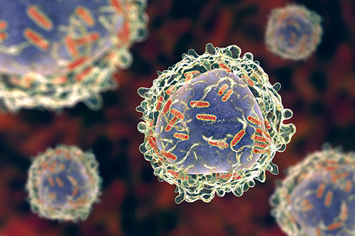
Studying Macrophages?
Bio-Rad offers a range of antibodies to detect macrophages, including markers specific for the M1 and M2 phenotypes.
References
Bustos R and Sobrino F (1992). Stimulation of glycolysis as an activation signal in rat peritoneal macrophages. Effect of glucocorticoids on this process. Biochem J 282, 299-303.
Hooftman A et al. (2023). Macrophage fumarate hydratase restrains mtRNA-mediated interferon production. Nature 615, 490-498.
Hou F et al. (2011). MAVS forms functional prion-like aggregates to activate and propagate antiviral innate immune response. Cell 146, 448-461.
Wang F et al. (2018). Glycolytic stimulation is not a requirement for M2 macrophage differentiation. Cell Metab 28, 463-475.
Zecchini V et al. (2023). Fumarate induces vesicular release of mtDNA to drive innate immunity. Nature 615, 499-506.
You may also be interested in...

We spoke to Professor Jon Austyn, Siamon’s first graduate student who carried out the original research identifying F4/80, about his remarkable scient...

Insights From Immunologists: Professor Jon Austyn
In this blog, we discuss new research from Meyer et al. (2022), suggesting that mild manipulation from T. gondii increases a gray wolf’s risk-taking b...
Puppetry of a Parasite: How Toxoplasma gondii Manipulates its Host
View more Immunology or Guest Blog blogs