Popular topics

Popular topics
-
References
Andrä I et al. (2020). An evaluation of T‐cell functionality after flow cytometry sorting revealed p38 MAPK activation. Cytometry Part A 97, 171-83.
Beliakova-Bethell N et al. (2014). The effect of cell subset isolation method on gene expression in leukocytes. Cytometry Part A 85, 94-104.
Llufrio EM et al. (2018). Sorting cells alters their redox state and cellular metabolome. Redox Biol 16, 381-387.
Richardson GM et al. (2015). Does FACS perturb gene expression. Cytometry Part A 87A, 166–175.
Zhou et al. (2016). Mitogen-activated protein kinases as key players in osmotic stress signalling. Biochim Biophys Acta 1860, 2037-52.
How to Avoid Cell Stress While Cell Sorting
It is important to avoid inducing stress responses when sorting cells, to be confident that any properties observed in downstream applications are not artefacts of the cell sorting process, and that cell viability and growth are not adversely affected. This blog describes the forces that cells undergo during sorting, and practical steps you can take to minimize cell stress and get great results in your downstream applications, like RNA sequencing.
Which Forces Do Cells Undergo During Sorting?
Cells are exposed to many stresses and forces during fluorescence-activated cell sorting. These forces have the potential to activate stress pathways, alter cellular structure and function, and affect cell metabolism (Llufrio et al. 2018). These effects could confound the results of experiments and should ideally be avoided.
During the short amount of time that a cell spends in a flow cytometer, it becomes exposed to high pressure and decompression, acceleration, high speed, shear forces, and laser illumination. When the cell exits the stream, it then experiences electrical charge and forces of impact, as well as rapid temperature change once it hits the collection tube (Andrä et al. 2020). All of these have the potential to result in changes in gene expression, cell health, and cell morphology.
How Does Cell Sorting Induce Stress?
One familiar consequence of cell stress is cell death. It can be very disappointing to spend time preparing cells and running them through a cell sorter only to notice that they are behaving oddly – or worse – that they are dead. By understanding what causes cell stress, you can make small changes to minimize the impact, keep your cells happy, and isolate pure populations of even rare cell types.
Various research groups have looked at the effect of sorting parameters on cell stress. While some studies have shown only a minimal impact of cell sorting on some parameters, like gene expression (Richardson et al. 2015, Beliakova-Bethell et al. 2014), others have seen significant effects on, for example, the metabolic profile (Llufrio et al. 2018).
The instrument used for cell sorting can also have an impact on cell stress due to the time spent in the system and the force used. Bio-Rad’s S3e Cell Sorter is designed to preserve cell viability and vitality as it is gentle even when sorting at high speeds. Researchers using the S3e Cell Sorter have discovered that cell sorting activated stress signaling through p38 MAP kinase (MAPK), but no functional or structural changes were observed in sorted versus unsorted cells. Patterns of gene transcription were also not significantly affected. This suggests that the S3e Cell Sorter is gentle on cells and strengthens its use upstream of RNA-Seq or other experiments (Andrä et al. 2020).
Practical Steps to Minimize Stress
While not every aspect of cell stress relating to sorting can be controlled – you probably can’t change the requirement for cells to move at relatively high speed, or to be subjected to electrostatic change and deflected into tubes - there are still some choices you can make to ensure your experiments are as successful and as stress-free as possible (for your cells!):
1) Optimize Your Buffer Solutions
Buffer solutions can affect cell stress by altering osmolarity or pH, or by adding traces of mitogens (Andrä et al. 2020). Osmotic stress, for example, causes cells to either swell or shrink and harms cellular activities via MAPK signaling (Zhou et al. 2016). Choosing the correct buffer solution before and after sorting is important. As a general rule, adding BSA or FBS to buffer solution will help to keep cells healthy.
An appropriate buffer will depend on your cell type, whether cells are usually adherent or happy in suspension, and whether or not your sample contains a high density of dead cells. It is tempting to think that culture media will be best for sorting cells, but it comes with downsides: pH regulation is suboptimal and the calcium chloride component may not be compatible with the instrument sort buffer.
2) Think about Impact and Charge
Cells may be damaged if they hit the side of a collection tube with a certain trajectory after sorting. The plastic in collection tubes has a negative charge, and a dry part of a tube can attract droplets containing cells that have been deflected using electrostatic charge. As the droplet evaporates, viability reduces and the cell may die. Some protocols recommend precoating collection tubes with buffer solution, masking the plastic’s charge and improving viability.
3) Check Your Pressure
Incredibly, cells can be propelled through a sorter at 44 mph or even faster: the speed will depend on the pressure. Running cells at higher speed or pressure can make your experiments faster, but could expose cells to more intense forces with detrimental effects on their biology. In addition, some cells (such as dendritic cells) just don’t perform well under pressure whereas others are relatively robust. Among other factors, any cells with structure on the outside of their membranes will be particularly likely to be damaged during sorting.
Generally, using a lower pressure can decrease the shear forces that cells experience. It also ensures that the core stream is narrow and has a consistent velocity. Wider, inconsistent streams can result in low cell recovery via a process known as “drop delay”.
Nozzle size is an important consideration relating to pressure – cell sorters come with a variety of nozzle sizes with specific pressure ranges. If you are sorting larger cells, you will need a wider nozzle. Inappropriate nozzle sizes can increase cell stress and decrease viability by causing inappropriate deposition and intense shear force.
4) Use the Best Temperature
As with any other molecular biology process, it is important to keep your cells at a healthy temperature. Generally, keeping cells on ice is advisable for slowing metabolic processes, but cold temperatures can also prevent cells from repairing any damage caused by the sorting process. Some mammalian cells will become stressed and die quickly at 4°C, so it is best to check the literature to find the best practice for your cell line.
5) Factor in Your Pre-Sort Treatments
Even if you optimize your pressure, temperature, and components of your buffer solutions, you might notice that cells are more stressed after some experiments than others, and recovery might differ between samples or experiments. This could be due to pre-sort experimental treatments that cells are exposed to.
For example, cells treated with drugs or cells that are apoptotic might be more damaged during the sort process than untreated and non-apoptotic cells. To understand whether this is affecting your experiment, you may want to do a comparable non-sorted experiment using cells treated with your different experimental conditions.
Could the S3e Cell Sorter Help You Minimize Cell Stress?
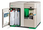 Not all cell sorters are built the same and the S3e Cell Sorter has been designed to help preserve cellular integrity without compromising power or efficiency. The sorter maintains a constant stream velocity during runs, avoiding the shear forces that are associated with rapid pressure changes within fluid streams. Instead, the S3e Cell Sorter uses jet-in-air sorting to preserve cell health and has a maximum sorting rate of 10 K events/sec, and can sort 12 million cells in 20 min.
References
Andrä I et al. (2020). An evaluation of T‐cell functionality after flow cytometry sorting revealed p38 MAPK activation. Cytometry Part A 97, 171-83.
Beliakova-Bethell N et al. (2014). The effect of cell subset isolation method on gene expression in leukocytes. Cytometry Part A 85, 94-104.
Llufrio EM et al. (2018). Sorting cells alters their redox state and cellular metabolome. Redox Biol 16, 381-387.
Richardson GM et al. (2015). Does FACS perturb gene expression. Cytometry Part A 87A, 166–175.
Zhou et al. (2016). Mitogen-activated protein kinases as key players in osmotic stress signalling. Biochim Biophys Acta 1860, 2037-52.
You may also be interested in...

This blog discusses some best practices for storing and using your antibodies to get the most out of them in your experiments....

Treat Them Right! – Best Practices for Storing and Working with Antibodies
In this blog, we cover some key considerations to keep in mind when presenting your data, with a particular focus on the differences between an extern...
Top Tips for Presenting Your Flow Data
View more Applications or Tips&Tricks blogs