Western Blot: Detection with Substrate
- On This Page
- Detection with substrate
s

s


Overview
Now that the target protein has been specifically tagged with an appropriately labeled antibody, and excess antibody has been washed away, the label will be used to identify the location of the target protein on the blot. Some labels can be detected immediately, without any further processing.
Fluorescent tags, for example, simply require the right equipment to observe and record the fluorescent signal. In Figure 12, a Western blot is probed with an anti-tubulin primary antibody and a DyLight® 800 conjugated secondary antibody.
Note here that the molecular weight standards at the far left are labeled to produce a red signal allowing them to be easily distinguished from the samples. This highlights one of the advantages of fluorescent labels, which is that multiple labels can be seen on the same blot in the same experiment.

Figure 12: Western Detection of Tubulin using DyLight® 800 secondary antibody
The most common antibody label used in Western blots is HrP, a small, stable enzyme with high specificity and rapid turnover. HrP is deactivated by sodium azide, so it is imperative that no azide is present in the blocking, dilution, or washing solutions. At Bio-Rad we offer a specialized buffer for the long term storage and dilution of HrP labeled antibodies, BUF052A, which can be used with all antibodies except those generated against rabbit immunoglobulins.
The HrP label is detected when it is exposed to a substrate solution in the final step of the immunodetection procedure. Substrate solutions for Western blotting are chemical reagents that are acted upon by the enzyme to yield a signal that can be easily measured. HrP label is typically detected with either colorimetric or chemiluminescent substrates.
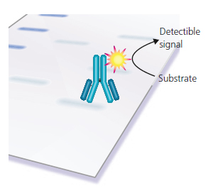
Figure 13: Detection of the Antibody Label. The antibody label is exposed to substrate in the final stages, creating a visible band either on the surface of the blot (colorimetric substrates), or as light emission (ECL substrate) captured on X-ray film or with a CCD camera.
Colorimetric substrates for HrP produce brown (DAB) or purple/black (4CN, TMB, NBT/BCIP) bands directly on the surface of the blot. These substrates are very easy to use and take from a few minutes to a few hours to produce visible bands. Detection limits for colorimetric substrates are in the low nanogram range.
The colorimetric detection reaction proceeds until stopped, leading to the risk of overdevelopment and less flexibility of results produced. The signal also tends to fade over time, so the record is less permanent.
More routinely, HrP is used with ECL (enhanced chemiluminescence) detection. For ECL detection, the substrate is luminol which is oxidized by HrP in the presence of H2O2 and an enhancer to produce light. The emitted light is detected by exposing the Western blot to X-ray film, or by using a CCD camera for light capture.
The emitted light forms a band on the film, or on the screen of the imaging system, indicating where the HrP-labeled antibody has bound to the target protein.
ECL detection of HrP is extraordinarily sensitive, allowing for the visualization of picogram to femtogram amounts of target protein. Furthermore, since multiple film or CCD exposures can be made with ease, little effort is required to produce a suitable permanent record for analysis or publication. Detection times for ECL substrates range from a few seconds to about 30 minutes and can usually be done or redone over a period of several hours.
Some molecular weight markers are designed to produce a signal during ECL detection yielding a visible ladder that is convenient for identifying the bands produced during immunodetection. A discussion of Western blotting results is found in the next chapter.
| Indirect vs. Direct Detection | Test Blots, Slot Blots, and Dot Blots |